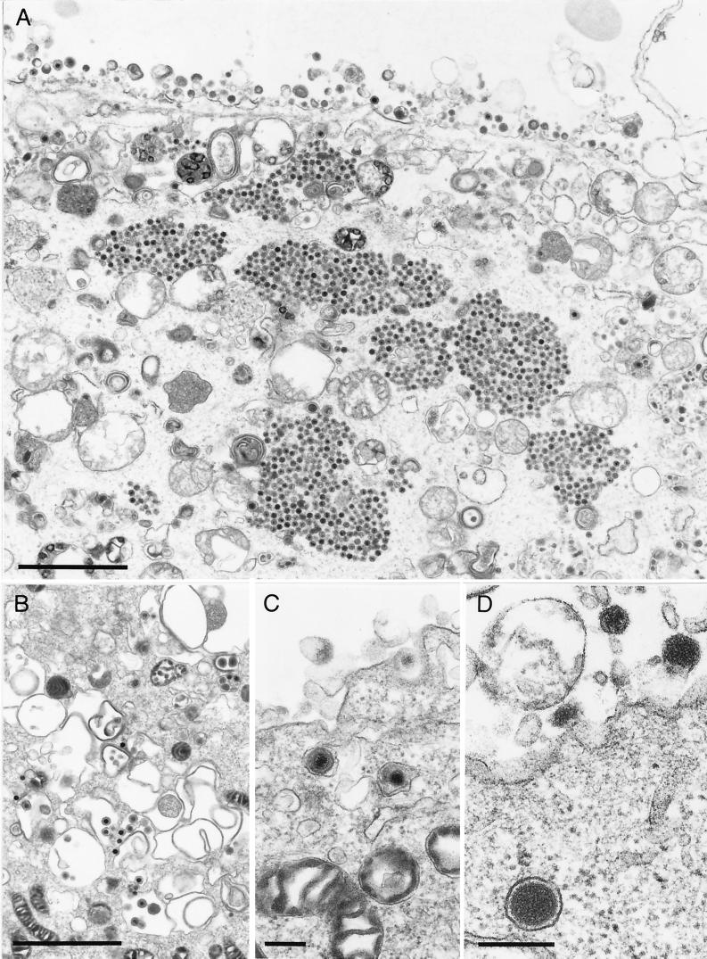
FIG. 6.
Production of L-particles in PrV-ΔUL37-infected RK13 cells. RK13 cells were infected as described in the legend to Fig. 5. (A) Overview demonstrating aggregation of intracytoplasmic capsids as well as numerous extracellular L particles. (B and C) Normal secondary envelopment (B) as well as enveloped virions in vesicles presumably during transport to the surface (C) were also observed, although rarely. (D) Particularly striking was the production of L particles (see also panel B). Bars in panels A and B and panels C and D are 2 μm and 250 nm, respectively.

