FIG. 7.
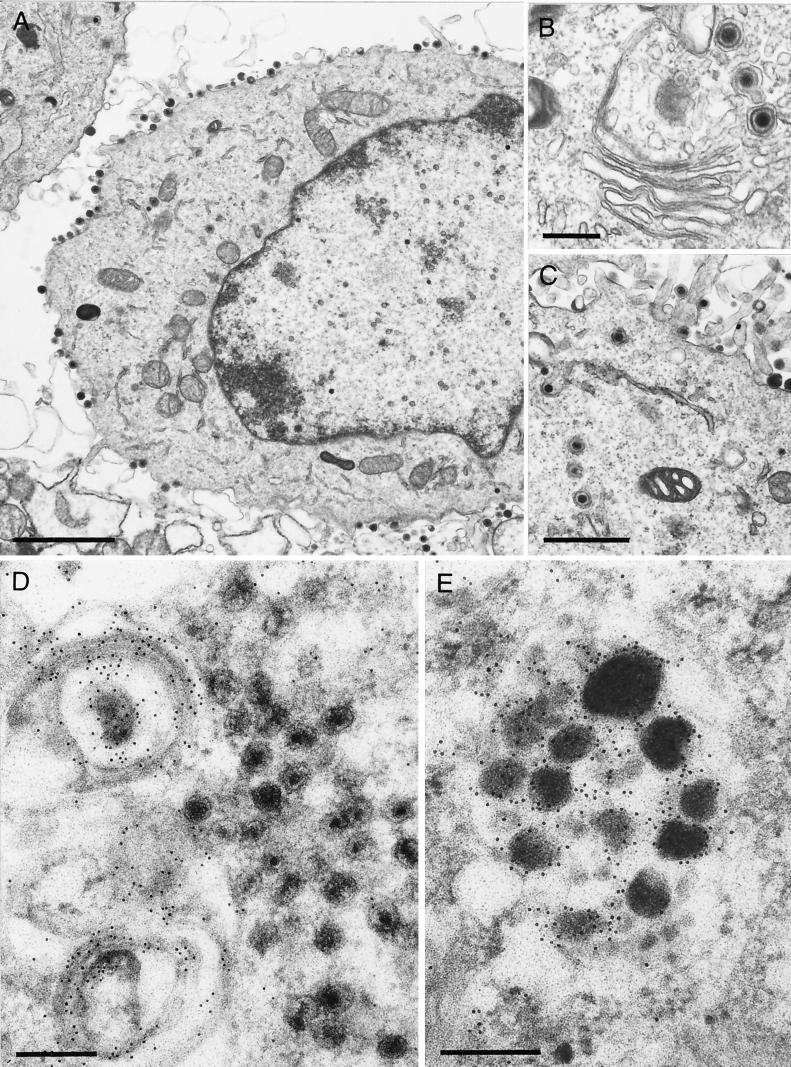
EM and immuno-EM of PrV-ΔUL37-infected RK13-UL37 and RK13 cells. (A to C) After infection of complementing RK13-UL37 cells with PrV-ΔUL37, all stages of normal virion maturation were observed (A), including unimpeded secondary envelopment (B) as well as transport to and accumulation at the plasma membrane (C). (D) To detect the major tegument protein UL49, PrV-ΔUL37-infected RK13 cells were labeled with a monospecific polyclonal anti-UL49 serum followed by incubation with gold-tagged secondary antibodies. Label was primarily detected in conjunction with vesicular membranes in the trans-Golgi area, the site of secondary envelopment. No label was detected in the accumulated capsids. (E) In contrast, L particles were heavily labeled by the anti-UL49 serum. Bars in panels A, B, C, and D and E are 2 μm, 0.5 μm, 1 μm, and 250 nm, respectively.

