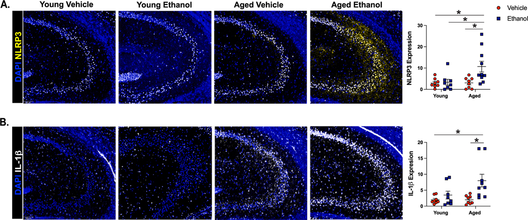
Fig. 2. Aged Mice Express Amplified Markers of NLRP3 Inflammasome Activation in the Hippocampus After Binge Ethanol Exposure Compared to Young.
Young (3–4 months old) and aged (18–20 months old) female C57BL/6N mice were exposed to intragastric gavages of ethanol (3 g/kg) or vehicle every other day for 10 total exposures. Multispectral imaging was performed on left hemispheres of young and aged ethanol and vehicle treated mice. Two sections from each brain hemisphere was imaged using a whole slice scanning system and representative images at 10x magnification are shown. Expression of percent positive area for (A) NLRP3 and (B) IL-1β in CA3 of the hippocampus were quantified using QuPath software; data are represented as the average of two sections/brain. N = 8–10 per group, means and SEM are reported. Values were significantly different from each other determined by 2-way ANOVA with Tukey’s post hoc test, ** p < 0.01, *** p < 0.001.

