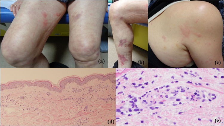
Figure 1.
Clinical and histopathological features. (a–c) Physical examination revealed wheals, edematous erythema, purpura, and livedo reticularis. (d) Histopathological examination of the edematous erythema showing capillary dilation and perivascular cellular infiltration. (HE staining, x40) (e) Histopathological examination of the edematous erythema showing perivascular neutrophil and eosinophil infiltration, nuclear dust, vessel wall destruction, and fibrin deposition in the upper dermis (HE staining, x400).

