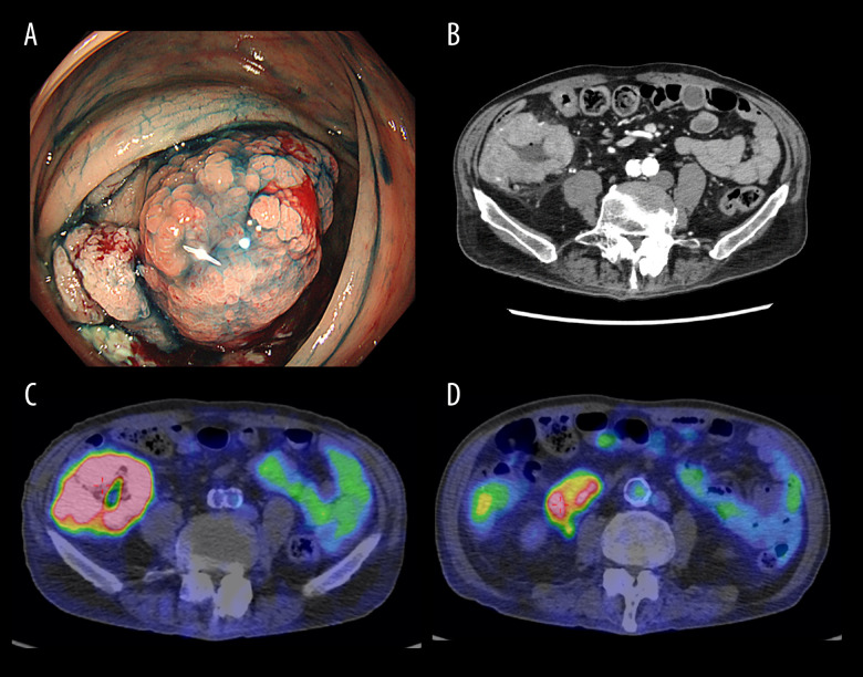
Figure 1.
(A) Hemorrhagic circumferential ulcer lesion in the ileocecal region. (B) Wall thickening mainly in the cecum. The serosal surface is irregular and fatty tissue density is elevated. There is a lymphadenopathy of less than 6 cm in length in the vicinity of the lesion. (C) SUVmax 13.9 in the primary lesion. There is also irregular wall thickening, suggesting a known mass lesion. (D) Lymph nodes also accumulated with SUV max 5.7.

