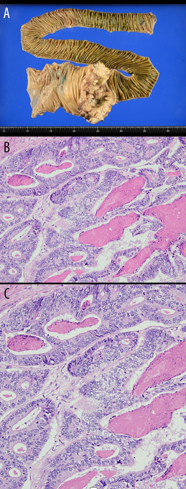
Figure 4.

Pathological examination. (A) Moderately differentiated tubular adenocarcinoma: tub 2 > tub 1. pT3(SS), lymph node; [#201(0/5), #202(4/6), #203(0/0)]. (B) Severe tumor necrosis. No bacteria were observed in the tissues. (C) A photomicrograph of the histopathology of the primary adenocarcinoma of the ileocecal region in a 72-year-old man with diabetes. The histopathology shows an adenocarcinoma with gland formation, cells with cytological atypia, mitoses, and areas of inflammation and necrosis, consistent with a moderate to high grade adenocarcinoma. Hematoxylin and eosin. Magnification × 40.
