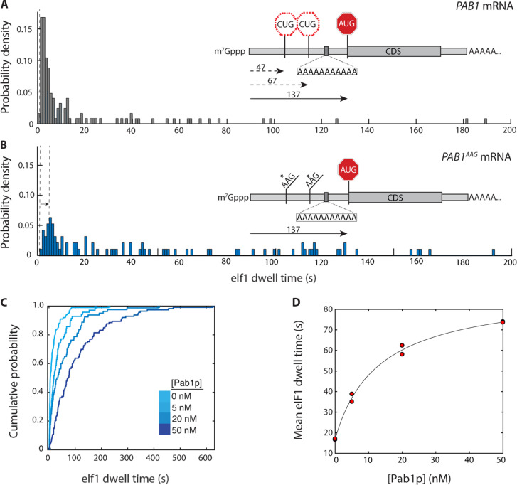
Fig. 4. Regulation of scanning by S. cerevisiae poly(A)-binding protein.
(A) eIF1 dwell-time distribution for the PAB1 mRNA (n = 125 molecules). Inset: Relative locations of upstream CUG triplets and PAB1 main-ORF AUG codon, and location of internal oligo(A) 11-mer. (B) eIF1 dwell-time distribution for the PAB1AAG mRNA, in which both upstream CUG triplets are substituted by AAG (n = 95). (C) eIF1 dwell-time cumulative distribution functions for scanning on the PAB1 mRNA in the presence of varying concentrations of the poly(A)-binding protein, Pab1p (5 nM, n = 110; 20 nM, n = 103; 50 nM, n = 112). (D) Dependence of mean eIF1 dwell time on PAB1 mRNA on the concentration of Pab1p, with hyperbolic fit (Kd 13 nM; 95% confidence intervals 1.1, 25.6 nM). Replicate data points at 0 and 50 nM Pab1p overlap. The time added to scanning at saturating Pab1p concentration is 72 s (95% confidence intervals: 64 to 79 s).

