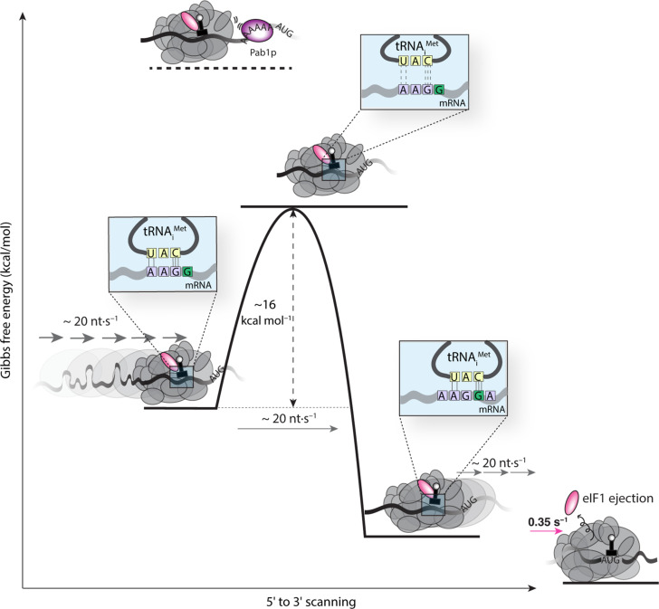
Fig. 5. Energetics of scanning.
The measured scanning rate of ~20 nt s−1 implies an energy barrier of ~16 kcal mol−1, on average, for PIC movement to the next nucleotide at each scanning step. The magnitude of this barrier corresponds to breakage of the hydrogen bonds in base-paired tRNA and mRNA; this base pairing by itself has an overall stability in the range of −3 kcal mol−1, with an enthalpic component of around −15 kcal mol−1. On the left of the schematic, one possible interaction during scanning includes five hydrogen bonds formed between a near-cognate mRNA triplet with a noncognate tRNA. Thermal fluctuation that disengages the base pair is expected to traverse an activation barrier equal to or exceeding the enthalpy of the hydrogen-bond interactions, as measured by our assay. Steric hindrance from Pablp binding to an oligo(A) site abutting the PIC increases this activation barrier, resulting in blocked scanning. eIF1 ejection stabilizes the PIC to the point where no further forward motion is possible.

