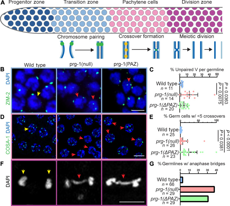
Fig. 3. Multiple meiotic processes are defective in prg-1 mutants.
(A) Schematic of meiotic progression and meiotic events by germ cell stage. Homolog pairing occurs in the TZ, followed by crossover formation in pachytene stage. Meiotic divisions occur in the division zone following diplotene stage. (B) Immunolocalization of ZIM-2 (green) in DAPI-stained nuclei (blue) of TZ cells. Two foci in a nucleus indicates unpaired homologs. One focus in a nucleus indicates paired homologs. Red arrowheads indicate nuclei with unpaired homologs. (C) Quantification of the percentage of germ cells with unpaired homologs per germline analyzed. (D) Localization of GFP::COSA-1 (green) in late pachytene nuclei stained with DAPI (blue). Red arrowheads indicate nuclei with fewer than five crossovers. (E) Quantification of the percentage of germ cells with less than five crossovers per nucleus per germline analyzed. (F) Representative DAPI (DNA, white) images of meiotically dividing germ cells from the division zone. Red arrowheads indicate anaphase bridges. (G) Quantification of percentage of germlines displaying meiotically dividing cells with anaphase bridges. Statistical significance was calculated by Student’s t test, mutant groups were compared to wild type. Scale bars, 5 μm. Each experiment was conducted at least in triplicate and over at least 25 to 30 germlines analyzed each time and at least 500 germ cells each time.

