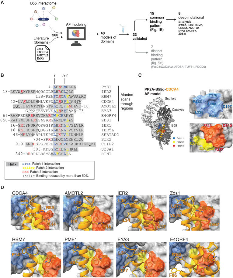
Fig. 1. Helical motifs engage a conserved binding pocket on PP2A-B55.
(A) Schematic of pipeline to identify PP2A-B55 binding elements. Generated with BioRender. (B) Alignment of validated instances with helices in gray and residues contacting patch 1 in blue, patch 2 in yellow, and patch 3 in red. The first eight proteins on the list were the proteins that were scanned by single alanine mutagenesis, and residues found to reduce binding by at least 50% upon mutation to alanine are in italic. (C) Model of the PP2A-B55-CDCA4 complex with the different patches in B55 indicated in different colors. (D) AF2 models of the indicated proteins and their interaction with B55. Core residues forming interactions in the different patches are presented as sticks. AF, AlphaFold.

