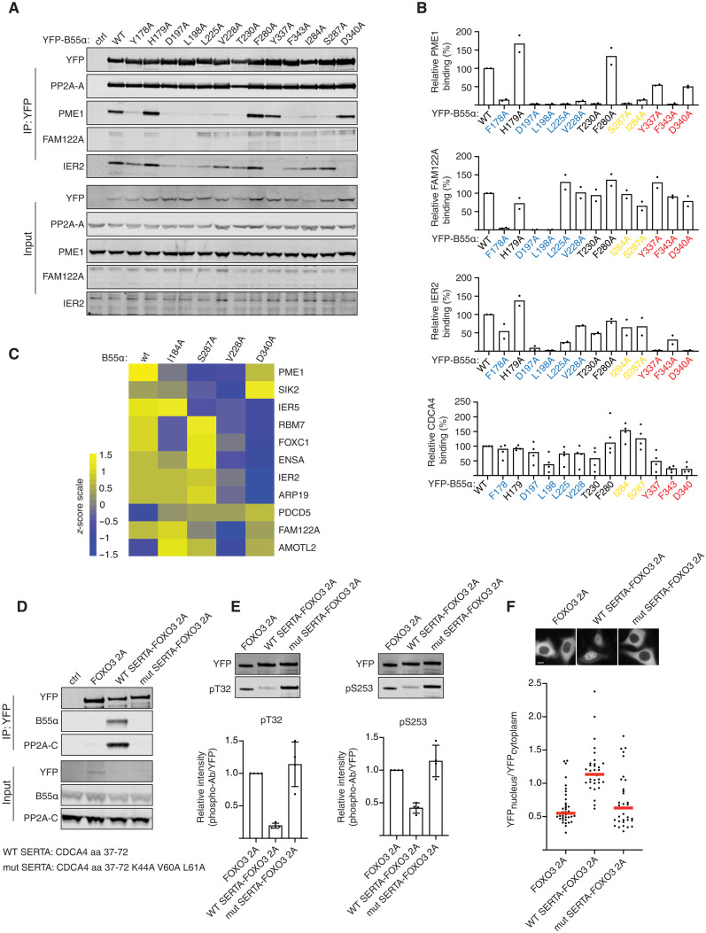
Fig. 2. Helical motifs can act as substrate specifying elements.
(A) Indicated B55 mutants were purified and binding to IER2, PME1, CDCA4, and FAM122A determined and quantified. (B) Quantifications of relative binding of indicated proteins to B55 of two or four biological replicates and the averages are shown. (C) Heatmap illustrating the changes in binding pattern of the indicated proteins to the different B55 variants as determined by MS. The scale is log 2. n = 4 technical replicates. (D) CDCA4 WT or mutant SERTA domain was fused to FOXO3 2A, and binding to PP2A-B55 was monitored by Western blot. (E) As in (D) but immunoprecipitations (IPs) probed with FOXO3 phospho-specific antibodies as indicated. n = 3 biological replicates. Error bars are mean with SD. (F) Subcellular localization of FOXO3 fusion proteins by live-cell microscopy. Shown is a representative experiment of three independent experiments. Each dot represents a cell analyzed. Median is indicated with red line. Scale bar, 10 μM.

