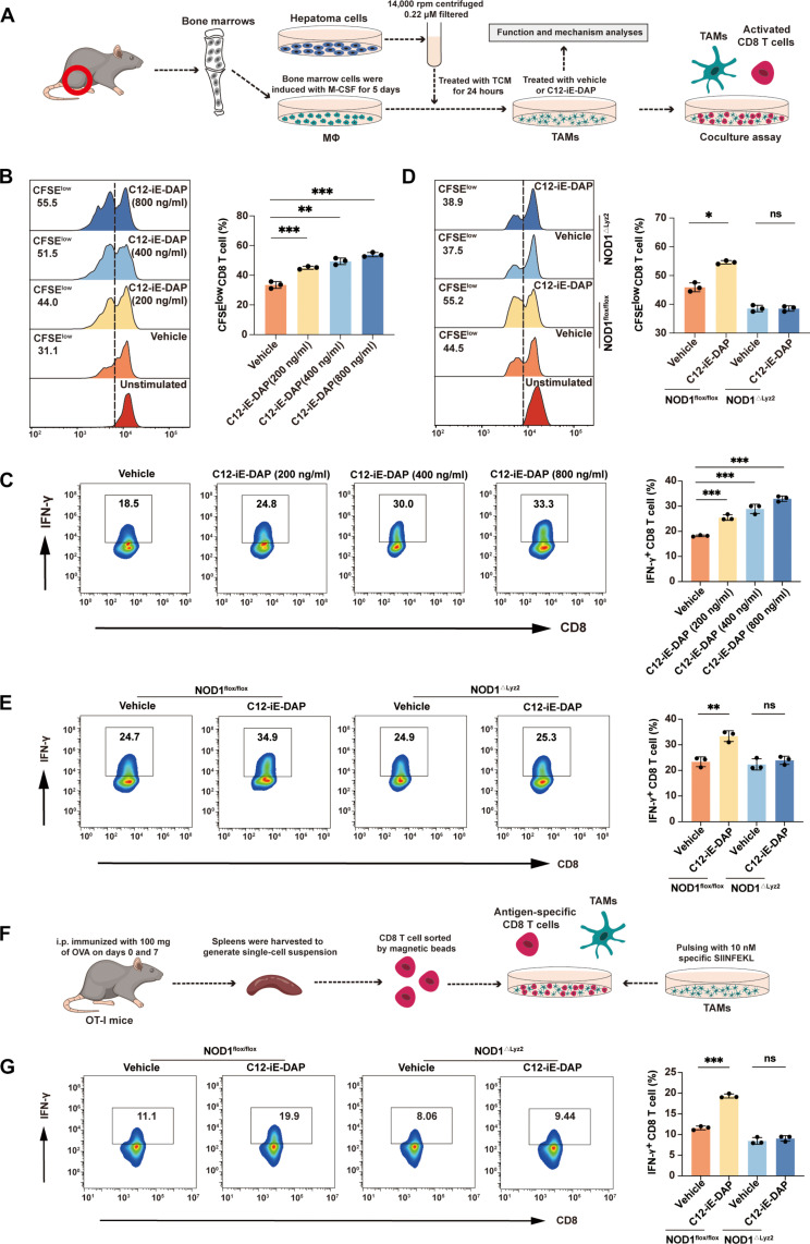
Fig. 2. NOD1 activation induces TAMs to acquire an immunostimulatory phenotype capable of supporting CD8+ T cell response.
(A) Schematic representation of the in vitro CD8+ T cell coculture assay. (B and D) Flow cytometric analysis of carboxyfluorescein diacetate succinimidyl ester (CFSE)–labeled CD8+ T cell proliferation after cocultured with TAMs that were generated from wild-type (WT) mice and treated with either vehicle or C12-iE-DAP (200, 400, and 800 ng/ml) for 24 hours (B) or from NOD1flox/flox or NOD1△Lyz2 mice and treated with either vehicle or C12-iE-DAP (400 ng/ml) for 24 hours (D). (C and E) Flow cytometry analysis of IFN-γ+ CD8+ T cells after cocultured with TAMs that were generated from WT mice and treated with vehicle or C12-iE-DAP (200, 400, and 800 ng/ml) for 24 hours (C) or from NOD1flox/flox or NOD1△Lyz2 mice and treated with either vehicle or C12-iE-DAP (400 ng/ml) for 24 hours (E). (F) Schematic representation of the in vitro antigen-specific CD8+ T cell coculture assay. i.p., intraperitoneal. (G) Flow cytometric analysis of IFN-γ+ OT-I CD8+ T cells after cocultured with TAMs that were generated from NOD1flox/flox or NOD1△Lyz2 mice and treated with either vehicle or C12-iE-DAP and then pulsed with SIINFEKL (OVA257-264). Statistical analysis was performed using the Student’s t test. Data were presented as mean with SD. ns, no significance; *P < 0.05, **P < 0.01, and ***P < 0.001. M-CSF, macrophage colony-stimulating factor.

