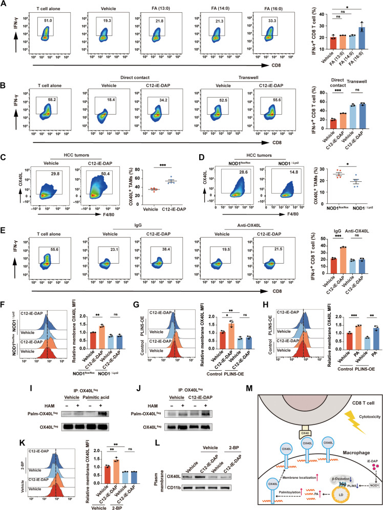
Fig. 5. NOD1-PLIN5–induced elevation of palmitic acid promotes macrophage OX40L membrane localization and activates CD8+ T cells.
(A) Flow cytometric analysis of IFN-γ+ CD8+ T cells cocultured with TAMs treated separately with 125 μM tridecanoic acid, myristic acid, or palmitic acid for 24 hours. (B) Flow cytometric analysis of IFN-γ+ CD8+ T cells cocultured with TAMs treated with either vehicle or C12-iE-DAP (400 ng/ml) in direct contact or separately in transwells. (C and D) Flow cytometric analysis of OX40L+ TAMs in orthotopic HCC tumors growing in WT mice treated intraperitoneally with either vehicle or C12-iE-DAP (C) or growing in NOD1flox/flox versus NOD1△Lyz2 mice (D). (E) Flow cytometric analysis of IFN-γ+ CD8+ T cells cocultured with TAMs pretreated with either immunoglobulin G (IgG) or anti-OX40L (5 μg/ml) and then treated with either vehicle or C12-iE-DAP (400 ng/ml). (F to H) Flow cytometric analysis of membrane OX40L in TAMs with different treatments. (I and J) OX40L palmitoylation was detected in TAMs using immunoprecipitation and acyl-biotin exchange (IP-ABE) assay. (K and L) Flow cytometric (K) and Western blot (L) analyses of membrane OX40L expression on TAMs pretreated with either vehicle or 2-bromopalmitate (2-BP; 50 μM), followed by treatment with either vehicle or C12-iE-DAP (400 ng/ml). (M) Schematic diagram depicting the regulatory role of NOD1 on TAMs. Statistical analysis was performed using the Student’s t test. Data were presented as mean with SD. *P < 0.05, **P < 0.01, and ***P < 0.001. MFI, mean fluorescence intensity; PA, palmitic acid; HAM, hydroxylamine.

