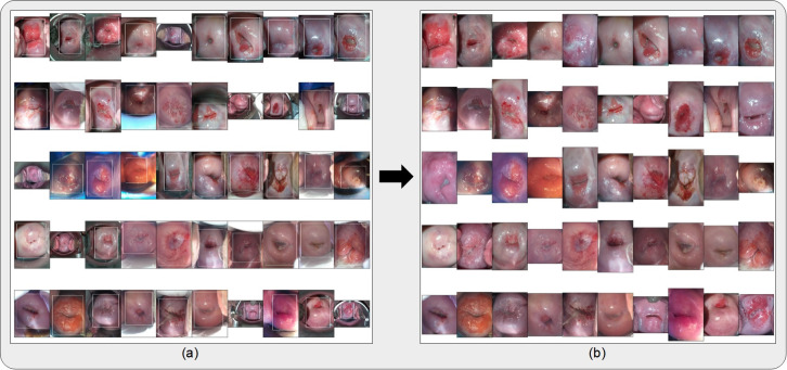
Fig 2.
(a) Bounding boxes generated from running the cervix detector, highlighted in white, around 50 randomly selected images from the external (“EXT”) dataset. The cervix detector utilized a YOLOv5 architecture trained on the “SEED” dataset images. (b) Bound and cropped images of the cervix which are passed onto the diagnostic classifier (AVE).

