
Figure 4. Microfluidic droplet devices for cell-laden microparticle generation.
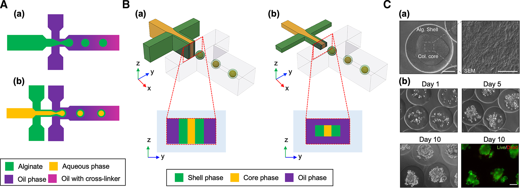
(A) Schematic of microfluidic droplet devices for generating (a) unstructured and (b) core-shell microparticles. (B) Schematic of (a) planar and (b) non-planar microfluidic droplet devices. (C) (a) Differential interference contrast (DIC) images of a collagen-alginate core-shell microparticle and scanning electron microscopy (SEM) images showing collagen fibers in the core. Scale bar: 100 μm for the DIC image and 5 μm for the SEM image. (b) Cell culture in collagen-alginate core-shell microparticles. Fluorescent image shows live (green)/dead (red) of the encapsulated MCF-7 cells. Scale bar: 100 μm. Reproduced with permission from ref. [38]. Copyright © 2017 American Chemical Society.