
Figure 1.
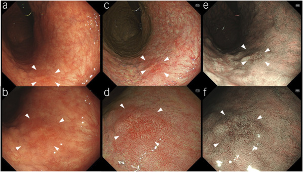
Representative images of target lesions. A depressed lesion in the middle third of the stomach is shown (arrowheads). The final histopathological diagnosis was well-differentiated adenocarcinoma confined to the mucosa. (a and b) On white-light imaging (WLI), the lesion appears as a reddish area with irregular margins and surfaces. (c and d) On texture and color enhancement imaging, the lesion appears as a reddish area with irregular margin surfaces, with a greater color difference than that in WLI. (e and f) Third-generation narrow-band imaging shows the lesion as a brownish area with irregular margins and surfaces.