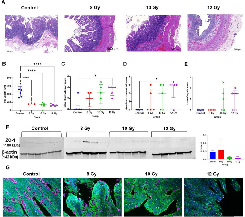
Fig. 2.

Radiation-induced alterations to the jejunum and epithelial barrier integrity. (A) Representative hematoxylin and eosin–stained images of jejunum from control and irradiated animals (20× objective, scale bar 500 μm). (B–E) Survivors are indicated with circles while decedents are represented by an x. (B) Villi length in μm. (C–E) Veterinary pathology scored metrics including villi blunting/fusion, mucosal apoptosis, and loss of crypts. Median ± interquartile range is indicated. (F) Western blots for protein expression of zonula occludens-1 (ZO-1) in jejunum tissue lysates in control and different irradiated dose group animals. Bars represent mean ± SD (G) Immunochemistry staining for ZO-1 expression (green), goblet cells using Wheat Germ Agglutinin (purple), and nuclei with DAPI (blue) (magnification, 20×). The color of each symbol represents the dose group. * P < 0.05, *** P < 0.0005, and **** P < 0.0001.
