FIGURE 4.

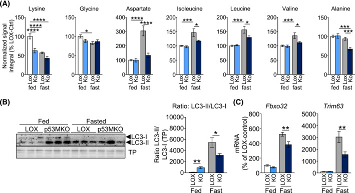
p53MKO mice demonstrate distinct changes in amino acid metabolism under fed and fasted conditions. (A) NMR spectroscopic analysis of intramuscular amino acids lysine, glycine, alanine, aspartate, isoleucine, leucine, valine and alanine in quadriceps of control (LOX) and p53MKO (KO) mice in the fed state (control: white bars; p53MKO: light blue bars) and following 16 h of food withdrawal (control: grey bars; p53MKO: dark blue bars). (B) Representative detection (left panel) of protein levels of LC3‐II and LC3‐I in quadriceps muscle of control and p53MKO mice in the fed state (LOX: white bars; p53MKO: blue bars) and following 16 h of food withdrawal (LOX: grey bars; p53MKO: dark blue bars) and quantification of LC3II:LC3I ratio normalized to total protein (right panel). (C) mRNA expression of Fbxo32 and Trim63 in quadriceps muscle under similar conditions. Results presented as mean ± SEM and compared by two‐way ANOVA (n = 5–10 for A and B, and n = 9–11 for C); *P < 0.05, **P < 0.01, ***P < 0.001, ****P < 0.0001.
