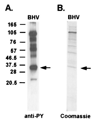
FIG. 1.
Tyrosine-phosphorylated BHV-1 structural proteins. (A) BHV-1 proteins were subjected to SDS-PAGE, immunoblotted, and probed with an antiphosphotyrosine antibody (PY54). Molecular mass markers are noted at the left in kilodaltons. (B) Coomassie blue staining of the purified BHV-1 structural proteins using the same sample preparation as that used for panel A. The protein band isolated and identified as VP22 is marked by arrows.

