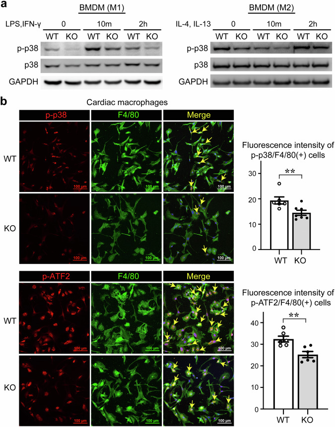
Fig. 5. IKKε knockout mice have lower levels of phosphorylated p38 in macrophages.
a Bone marrow-derived macrophages (BMDMs) were stimulated with 100 ng/mL LPS, 30 ng/mL IFN-γ, 30 ng/mL IL-4, and 30 ng/mL IL-13 for the indicated times. The protein levels of p-p38 and p38 in BMDMs were assessed in WT and IKKε KO mice using Western blotting. b Cardiac macrophages were isolated from heart tissues 3 days after myocardial infarction and immunofluorescence staining revealed the phosphorylation of p38 and its substrate ATF2. The fluorescence intensity indicating the phosphorylation of p38 and ATF2 was quantified in both the WT and IKKε KO groups. Scale bars: 100 μm. The data are presented as the means ± SEMs. **P < 0.01 (Student’s t-test).

