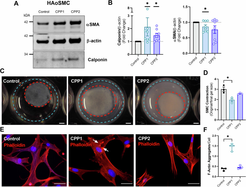
Fig. 3. Calciprotein particles decrease VSMC contractility and disorganize F-actin network.
Human aortic vascular smooth muscle cells were incubated with CPP1 or CPP2 for 24 h to study the effect of CPPs on VSMC function/contractility. A, B Representative image of western blot of αSMA, β-actin and calponin in HAoSMCs treated for 24 h with CPP1 or CPP2, including analysis graphs. C, D Representative images and analysis of collagen contraction assay after CPP incubation. Scale bar = 2 mm. E, F Representative images and analysis of phalloidin-stained HAoSMCs after 24 h incubation with CPPs to visualize F-actin. White arrows indicate F-actin aggregates. Scale bar = 40 µm. Statistical analyses: B One-sample T test on Log2 transformed data (hypothetical value = 0). n = 8. D, F Kruskal–Wallis test with Dunn’s post hoc test for multiple comparisons. n = 3. *p < 0.05. Error bars represent standard error of the mean (SEM). PE Phenylephrine, (HAo)SMC (Human aortic) smooth muscle cell, CPP1 Primary calciprotein particle, CPP2 Secondary calciprotein particle.

