Figure 5.

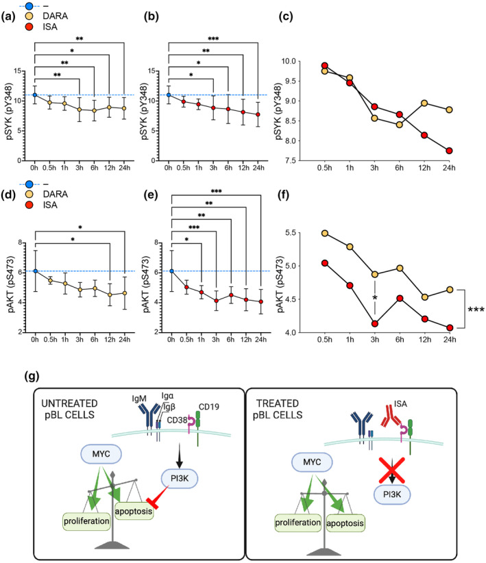
Anti‐CD38 mAbs impair PI3K pathway signalling in pBL cells. (a, b) Time‐course analysis of SYK phosphorylation levels post anti‐CD38 mAb treatment over 24 h. (c) Comparative kinetic analysis of SYK dephosphorylation between DARA and ISA‐treated cells. (d, e) The phosphorylation status of AKT over a 24‐h period post anti‐CD38 mAb treatment. (f) Differential analysis of pAKT level kinetics between DARA and ISA treatments. Statistical significance was assessed using one‐way ANOVA with the post hoc Tukey test comparing untreated vs treated, indicated by: *P < 0.05; **P < 0.01; ***P < 0.001. In (c) and (f), the statistical significance was assessed using the unpaired t‐test for the comparison between DARA vs ISA in each time point and the paired t‐test for the comparison of the whole kinetic, and indicated by *P < 0.05; ***P < 0.001. Data are presented as mean ± SD. Non‐significance is not denoted. Data are representative of two independent experiments. All results were normalised and merged for consistent interpretation. (g) Schematic representation of the proposed effect of anti‐CD38 mAb (ISA) treatment on MYC/PI3K pathways and consequences on the proliferation/apoptosis balance.
