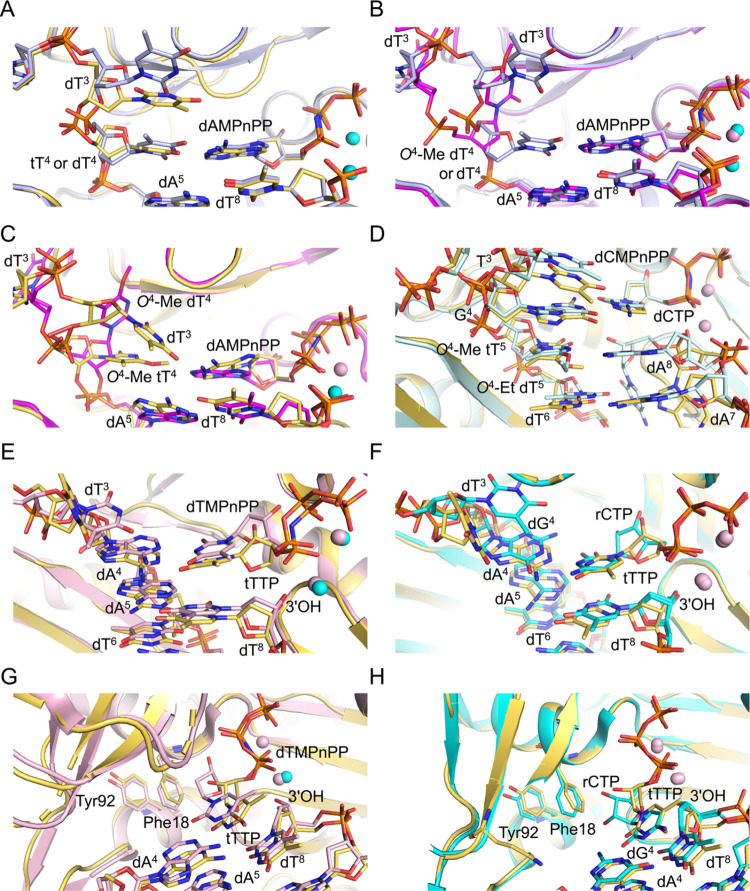
Figure 6.
Overlays of crystal structures of ternary hPol η complexes with all-DNA or TNA-modified template strands with thymine or O4-Me or -Et adducted thymine nucleobases. (A) Comparison between insertion complexes featuring either template tT4 (yellow carbons, this work) or dT4 (gray carbons, PDB ID 3MR2(42)) opposite incoming dAMPnPP. (B) Comparison between insertion complexes featuring either template dT4 (gray carbons, PDB ID 3MR2) or O4-Me dT4 (magenta carbons, PDB ID 5DLF(41)) opposite incoming dAMPnPP. (C) Comparison between insertion complexes featuring O4-Me tT4 (yellow carbons, in this work) or O4-Me dT4 (magenta carbons, PDB ID 5DLF) opposite incoming dAMPnPP. (D) Comparison between extension complexes featuring O4-Me tT5 (yellow carbons, this work) or O4-Et dT5 (light blue, PDB ID 5DQI(41)) paired opposite primer dA8 at the −1 postreplicative position and wedged between the replicating dG4:dCMPnPP (dCTP) and −2 position dT6:dA7 base pairs. (E) Comparison between ternary complexes of hPol η with incoming dTTP (light pink carbons, PDB ID 6PL7) and tTTP (yellow carbons, this work) opposite dA4 in the DNA template-primer duplex. (F) Comparison between ternary complexes of hPol η with incoming rCTP opposite dG4 (cyan carbons, PDB ID 5EWE(57)) or tTTP opposite dA4 (yellow carbons, this work). Coordinated Mg2+ and Ca2+ ions are depicted as cyan and pink spheres, respectively, in all of the figure panels. (G) Active-site view illustrating differences in the orientations of 2′-deoxyribose and threose sugar rings with respect to the Phe18 (steric gate) and Tyr92 (second line of defense) hPol η residues in the structures of complexes with dA:dTMPnPP (light pink carbons, PDB ID 6PL7) or dATP:tTTP (yellow carbons, this work), respectively. (H) Active-site view illustrating differences in the orientations of ribose and threose sugar rings with respect to the Phe18 and Tyr92 in the structures of complexes with dG:rCTP/dA:rCTP (cyan carbons, PDB ID 5EWE/6PL757) or dA:tTTP (yellow carbons, this work), respectively.

