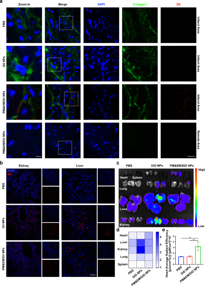
Figure 4.
PM&EM-NP-mediated delivery to the heart failure mice and biodistribution in other organs. (a) CLSM images of different DiI-labeled nanoparticles at fibrotic sites of the heart in the heart failure mice at 24 h postintravenous injection, scale bar = 5 μm, 20 μm. (b) CLSM images of different DiI-labeled nanoparticles in kidney and liver of the heart failure mice at 24 h postintravenous injection, scale bar = 15 μm. (c) Representative ex vivo fluorescence images of DiD fluorescence dye accumulated in different organs at 24 h postintravenous injection of DiD-labeled nanoparticles. (d) Heatmap of DiD fluorescence dye in different organs (n = 3). (e) Quantitative data of DiD fluorescence dye in the heart (mean ± SD, n = 3) **p < 0.01, one-way ANOVA, Tukey’s multiple comparison test.

