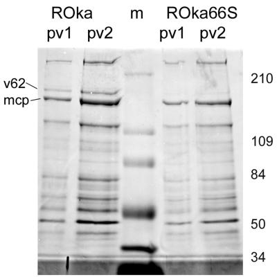
FIG. 3.
Coomassie blue-stained SDS-PAGE-separated polypeptide profile of two separate preparations of virions obtained from cells infected with either ROka or ROka66S. All preparations were derived following two sequential Ficoll gradient fractionations of infected-cell cytoplasmic extracts. The lane marked “m” represents molecular weight markers used to identify the sizes of the virion polypeptides, which are shown to the right in thousands. The 155-kDa major capsid protein (mcp) and the virion 175-kDa polypeptide (v62) are indicated on the left of the figure.

