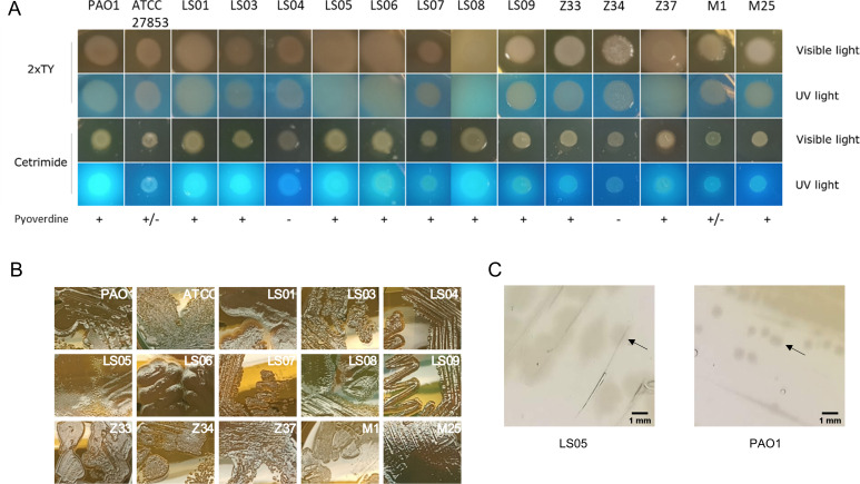
Fig 1.
Growth phenotype of clinical strains. (A) Colony morphology assay. Growth and pyoverdine production were analyzed by growing P. aeruginosa strains in cetrimide and 2xTY agar plates. Pictures were taken under UV and visible light after incubation at 37°C for 24 h. (B) Growth phenotype in 2xTY agar. The strains were inoculated as streaks in agar plates and incubated at 37°C for 24 h. (C) Comparison of colony size between LS05 and PAO1 strains in cetrimide agar. Colonies are indicated with black arrows.

