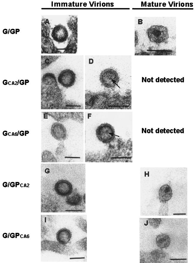
FIG. 7.
Electron microscopic analysis of the mutant viral particles. 293T cells were cotransfected with G/GP (A and B), GCA2/GP (C and D), GCA6/GP (E and F), GCA2/GP (C and D), and GCA6/GP (E and F). DNA constructs were harvested at 36 h posttransfection and processed for TSEM as described in Materials and Methods. Both immature (A, G, and I) and mature (B, H, and J) forms of the virus were detected on or near the surface of the cells transfected with G/GP (control), G/GPCA2, or G/GPCA6. Only immature virions were observed within the GCA2/GP (C and D) and GCA6/GP (E and F) preparations. (D and F) At times, an internal structure (arrow) within the inner ring of the immature virion was seen. Two diameters were measured for each viral particle: the longest and shortest diameters, roughly at right angles; the average of the two values was then taken as the diameter of the viral particle. Bars, 100 nm.

