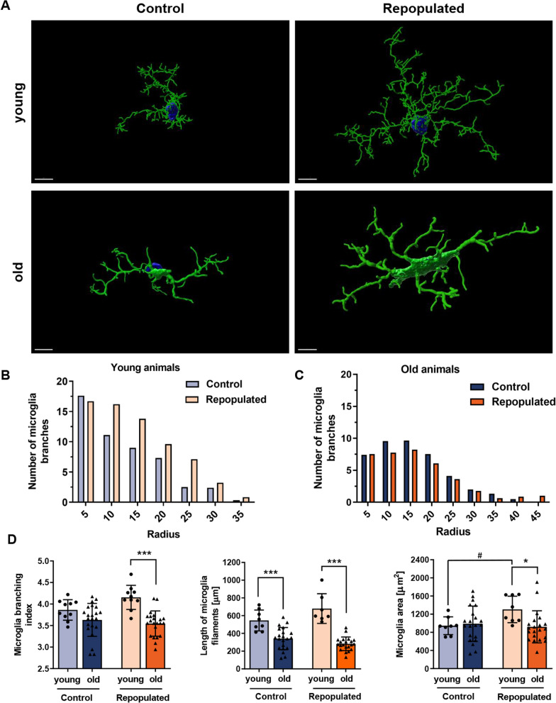
Fig. 5.
Different morphology of repopulated microglia from aged mice. A Representative confocal microscopy images of TMEM119+ MG. 3D models were created with the Imaris Software (Oxford Instruments, UK). Scale bar 7 µm. Notably, in young mice (3 months, n = 6) repopulated MG are more branched, with numerous elongated filaments with secondary processes located distal to the cell body. MG after repopulation have larger cell bodies. In old mice (18–22 months, n = 7) the repopulated MG are branched similarly to those in control mice and had fewer branches/processes than in the brains of young mice. B, C Quantification of control or repopulated MG branching with a Sholl analysis (radius 5 µm) in young (B) and old (C) mice, respectively. D Comparison of the MG branching (branching index and length of filaments) in brains of young and old mice (for branching, Sidak’s Test df = 60, p = 0.0001, for the length of MG filaments, Sidak’s Test control group df = 53, p = 0,0001, repopulated group df = 53, p = 0,0001); and the area covered by TMEM119+ MG (Sidak’s Test df = 54, p = 0.0193)

