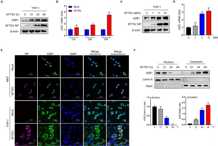
Fig 1.
HnRNP A2B1 is upregulated and translocated under SFTSV infection. (A) THP-1 cells were infected with SFTSV at a multiplicity of infection (MOI) of 10 for 12, 24, or 48 h. A2B1 protein levels were analyzed with Western blot. (B) THP-1 cells were infected with SFTSV at an MOI of 10 for 12, 24, or 48 h. A2B1 mRNA levels were analyzed with RT-qPCR. (C) THP-1 cells were infected with SFTSV at an MOI of 0, 1, 5, or 10 for 48 h. A2B1 protein levels were analyzed with Western blot. (D) THP-1 cells were infected with SFTSV at an MOI of 0, 1, 5, or 10 for 48 h. A2B1 mRNA levels were analyzed with RT-qPCR. (E) MEF cells and THP-1 cells were infected with SFTSV at an MOI of 10 or 0 (Mock control) for 48 h, SFTSV NP (purple), hnRNP A2B1 (green), and DAPI (blue) were analyzed with confocal microscopy. (F) MEF cells were infected with SFTSV at an MOI of 10 for the indicated time points. Nuclear and cytoplasmic proteins were separated. hnRNP A2B1, Lamin A, and Rab5 protein levels were analyzed with Western blot. Lamin A and Rab5 were nuclear and cytoplasmic index proteins, respectively. Nuclear and cytoplasmic Western blot data were semi-quantified and normalized against Lamin A and Rab5 protein loading control, respectively. Data were obtained from three independent experiments (n = 3). **P < 0.01, ***P < 0.001, ****P < 0.0001, ns, not significant.

