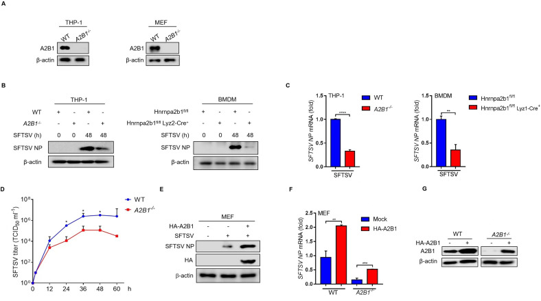
Fig 2.
HnRNP A2B1 upregulates SFTSV replication. (A) Knockout of hnRNP A2B1 in THP-1 cells and MEF cells was confirmed with Western blot. (B) BMDM cells were isolated from A2B1fl/fl and A2B1fl/flLyz2-Cre–/– mice. WT and A2B1-/- THP-1 and BMDM cells were infected with SFTSV at an MOI of 10 for 48 h, and SFTSV NP protein levels were analyzed with Western blot. (C) WT and A2B1-/- THP-1 and BMDM cells were infected with SFTSV at an MOI of 10 for 48 h, and SFTSV NP mRNA levels were analyzed with RT-qPCR. (D) WT and A2B1-/- MEF cells were infected with SFTSV for the indicated time points. SFTSV titers in cell culture supernatant were measured with immunofluorescence assay. (E) MEF cells were transfected with HA-tagged A2B1 for 24 h and then were infected with SFTSV at an MOI of 10 for 48 h. SFTSV NP protein levels were analyzed with Western blot. (F) WT and A2B1-/- MEF cells were transfected with HA-tagged A2B1 for 24 h, and then were infected with SFTSV at an MOI of 10 for 48 h. and SFTSV NP mRNA levels were analyzed with RT-qPCR. (G) WT and A2B1-/- MEF cells were transfected with HA-tagged A2B1 for 24 h, A2B1 protein levels were analyzed with Western blot. Data were obtained from three independent experiments (n = 3). **P < 0.01, ***P < 0.001, ****P < 0.0001, ns, not significant.

