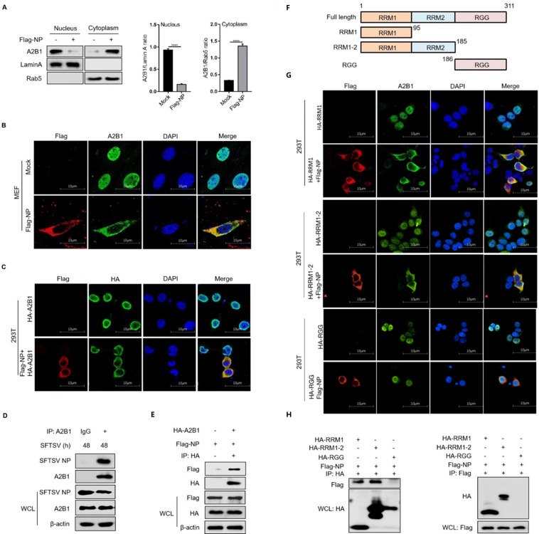
Fig 5.
SFTSV NP is important for the translocation of hnRNP A2B1. (A) MEF cells were transfected with Flag-tagged SFTSV NP for 24 h, nuclear and cytoplasmic proteins were separated, and hnRNP A2B1, Lamin A, and Rab5 protein levels were analyzed with Western blot. Western blot data were semi-quantified and normalized against Lamin A and Rab5 protein loading control, respectively. (B) MEF cells were transfected with Flag-tagged SFTSV NP for 24 h, Flag-tagged SFTSV NP (red), A2B1 (green), and DAPI (blue) were analyzed with confocal microscopy. (C) HEK293T cells were transfected with the indicated plasmids for 24 h, Flag-tagged SFTSV NP (red), HA-tagged A2B1 (green), and DAPI (blue) were analyzed with confocal microscopy. (D) THP-1 cells were infected with SFTSV at an MOI of 10 for 48 h, and interaction between SFTSV NP and A2B1 in THP-1 cells was analyzed with CO-IP. (E) HEK293T cells were transfected with HA-tagged A2B1, Flag-tagged SFTSV NP for 24 h, and interaction between Flag-tagged NP and HA-tagged A2B1 was detected with CO-IP. (F) The diagram of hnRNP A2B1 domains and the truncated hnRNP A2B1 construction. (G) HEK293T cells were transfected with the indicated plasmids for 24 h, Flag-tagged SFTSV NP (red), HA-tagged hnRNP A2B1 domains (green), and DAPI (blue) were analyzed with confocal microscopy. (H) HEK293T cells were transfected with the indicated plasmids for 24 h, and interaction between Flag-tagged NP and HA-tagged hnRNP A2B1 domains was detected by CO-IP. Data were obtained from three independent experiments (n = 3). ****P < 0.0001.

