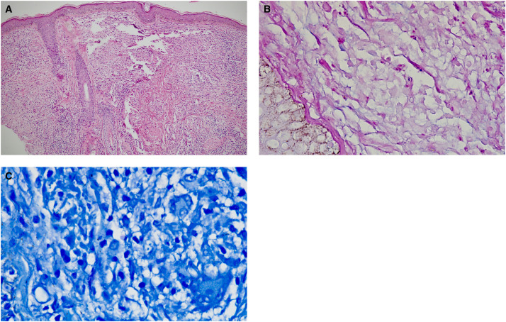
Figure 3.
(A and B) Skin biopsies showed presence of Grenz zone and band-like infiltrate in papillary dermis. Dermis showed mixed inflammatory infiltrates comprising of lymphocytes, histiocytes, plasma cells, and neutrophils. There was presence of well-formed epithelioid cell granulomas with scattered multinucleated giant cells. The histiocytes showed intractoplasmic amastigote forms of leishmania (hematoxylin and eosin stain, 40× and 100×) (C) Giemsa stain showed presence of Leishmanin–Donovan bodies (100×).

