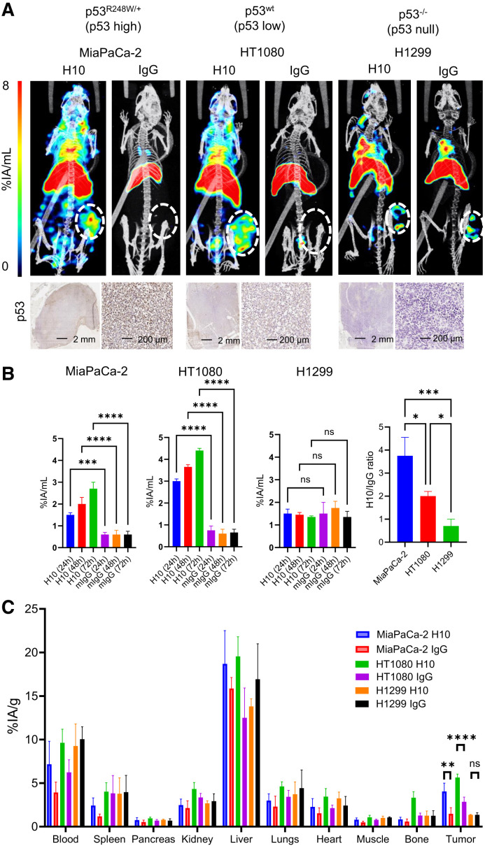
FIGURE 3.
(A) SPECT/CT imaging 72 h after intravenous administration of 111In-H10-TAT or 111In-mIgG-TAT (5 MBq, 5 μg) in BALB/c nu/nu mice bearing p53mut MiaPaCa-2, p53wt HT1080, or p53null H1299 xenografts. At bottom, immunohistochemistry staining for p53 in tissue harvested from tumor xenografts confirms p53 expression levels. (B–E) Volume-of-interest analysis of tumor uptake over time of 111In-H10-TAT or 111In-mIgG-TAT in mice bearing MiaPaCa-2, HT1080, or H1299 xenografts. Tumor uptake ratio of 111In-H10-TAT or 111In-mIgG-TAT is in line with p53 expression in tumor xenografts. (F) Full biodistribution data 72 h after intravenous administration of 111In-H10-TAT or 111In-mIgG-TAT shows differences only in tumor xenografts, not in normal tissue. *P < 0.05. **P < 0.01. ***P < 0.001. ****P < 0.0001. %IA = percentage injected activity; ns = not statistically significant.

