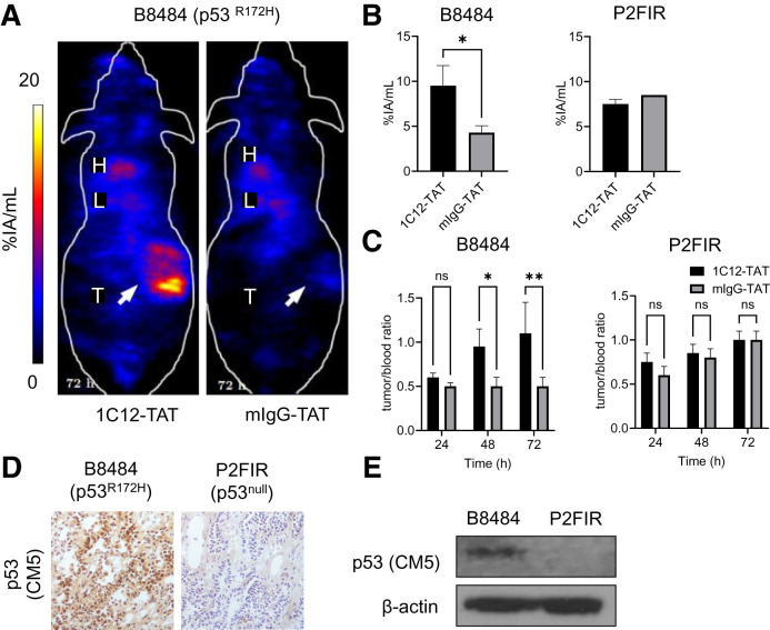
FIGURE 4.
(A) SPECT/CT imaging 72 h after intravenous administration of 111In-1C12-TAT vs. 111In-mIgG-TAT (5 MBq, 5 μg) in mice bearing B8484 murine PDAC allografts shows uptake of former but not latter. (B and C) Volume-of-interest analysis of tumor uptake, and tumor-to-blood ratios of 111In-1C12-TAT or 111In-mIgG-TAT in mice bearing p53R172H B8484 or p53null P2FIR allografts, show significant uptake in former, not latter. (D and E) Immunochemistry and Western blot analysis of p53 expression in tumor allograft tissue using rabbit polyclonal anti-p53 antibody CM5 confirmed p53 expression. *P < 0.05. **P < 0.01. %IA = percentage injected activity; H = heart; L = liver; ns = not statistically significant; T = tumor.

