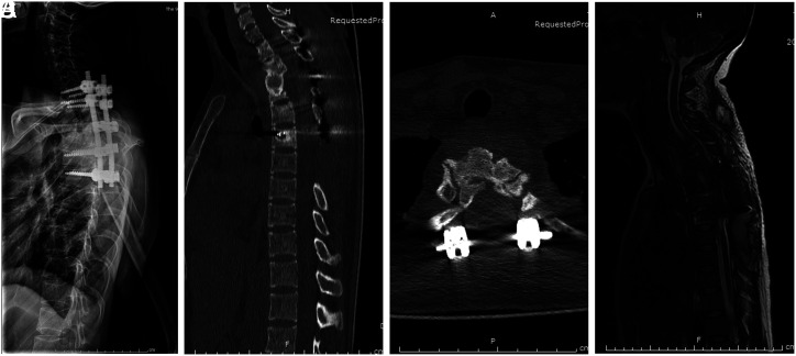
Figure 2.
(A) Postoperative lateral X-ray film, local kyphosis had improved significantly. (B) Postoperative CT, solid bone fusion in diseased segments. The local kyphotic Cobb angle was 16°. (C) Axial CT showed that the intervertebral bone graft had achieved bone fusion. (D) Postoperative MRI, spinal cord compression wholly relieved.

 Content of this journal is licensed under a