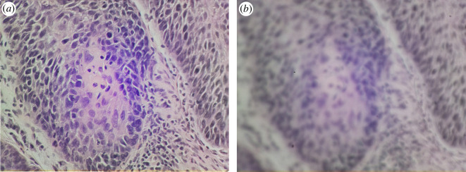
Figure 3.
Images of a squamous cell carcinoma from a vocal cord sample, captured on the OpenFlexure Microscope with a 40 objective. Panel (a) shows a focused image with well-aligned optics, while panel (b) shows the same area out of focus and with misaligned illumination. A trained user can distinguish between these images, refusing to give a diagnosis based on poor data.

