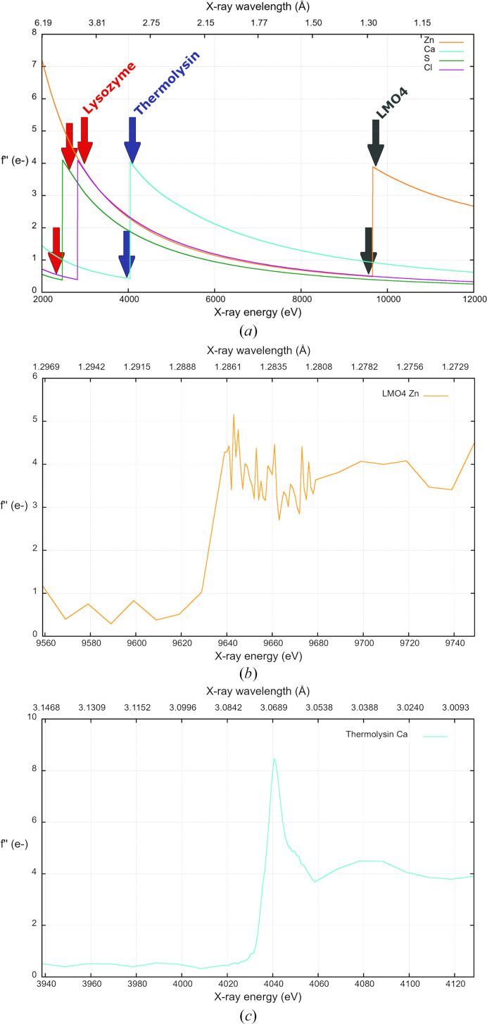
Figure 1.
Variation of f′′ with X-ray wavelength (or energy) showing absorption K edges for sulfur (green), chlorine (magenta), calcium (cyan) and zinc (orange). (a) Theoretical f′′ values obtained from the website https://skuld.bmsc.washington.edu/scatter/. Wavelengths used for data collections are marked with red, blue and grey arrows for lysozyme, thermolysin and LMO4, respectively. (b) Zn K-edge absorption-edge scan as determined by CHOOCH (Evans & Pettifer, 2001 ▸) measured from an LMO4 crystal. (c) Ca K-edge absorption edge scan as determined by CHOOCH and measured from a thermolysin crystal.

