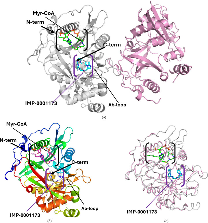
Figure 1.
PvNMT structure. (a) There are two PvNMT monomers in the asymmetric unit. Chain A (gray) has a bound Myr-CoA (green sticks) and inhibitor IMP-0001173 (blue sticks). Chain B (pink) shows a PvNMT monomer in the apo state. (b) Cartoon of PvNMT monomer A colored in a rainbow from blue at the N-terminus to red at the C-terminus. Myr-CoA (magenta sticks) and the inhibitor IMP-0001173 (white sticks) are shown. (c) Superposition of the monomers chain A (with ligands, gray) and chain B (apo PvNMT, pink). The substrate-binding cavity is indicated in purple parentheses, while the Myr-CoA-binding cavity is shown in black parentheses.

