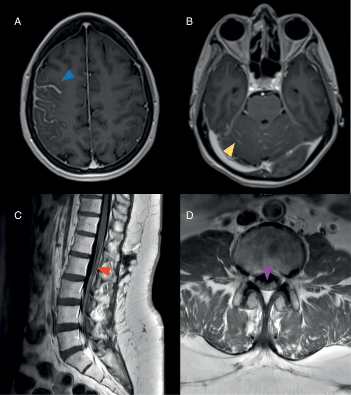
Figure 2.
Classic magnetic resonance imaging (MRI) features in patients with leptomeningeal metastases. (A) Thick sulcal enhancement involving the right frontoparietal sulci (arrowhead) on MRI brain axial T1-MPRAGE post-contrast images. (B) Linear ill-defined enhancement and nodules studding the cerebellar folia (arrowhead) on MRI brain axial T1-MPRAGE post-contrast images. Smooth leptomeningeal enhancement may also be seen coating the cranial nerves in the posterior fossa. (C) Smooth widespread enhancement of the conus medullaris and cauda equina (arrowhead) on MRI lumbar spine sagittal T1 post-contrast images. (D) Enhancement and clumping of the cauda equina nerve roots (arrowhead) on MRI lumbar spine axial T1 post-contrast images.

