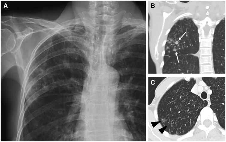
Figure 3.
Pulmonary tuberculous nodules in a 61-year-old woman. (A) Frontal chest radiograph and (B) coronal CT image show reticulonodular infiltration in the right upper lobe, with several nodules (white arrows). (C) Axial CT image shows tree-in-bud opacities in central zone as well as in the peripheral posterior aspect of the lung (black arrowheads).

