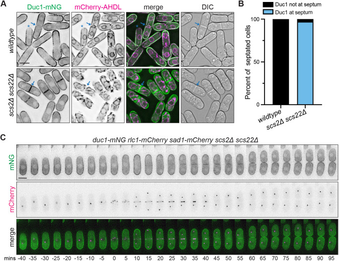
Fig. 2.
Duc1 is a PM protein that is excluded from the cell division site. (A) Live-cell imaging of Duc1–mNG and mCherry–AHDL in wild-type or scs2Δ scs22Δ cells. Blue arrows mark a septum, and blue asterisks mark a cell tip where localization is excluded. (B) Quantification of the frequency of Duc1 localization to the septum in septated cells from A. n=50 cells for each from two independent replicates. (C) Live-cell time-lapse imaging of duc1-mNG rlc1-mCherry sad1-mcherry scs2Δ scs22Δ. Elapsed time is in minutes. Images were acquired every 5 min and time zero represents the first frame of SPB separation. Images are representative of two biological replicates. DIC, differential interference contrast image. Scale bars: 5 µm.

