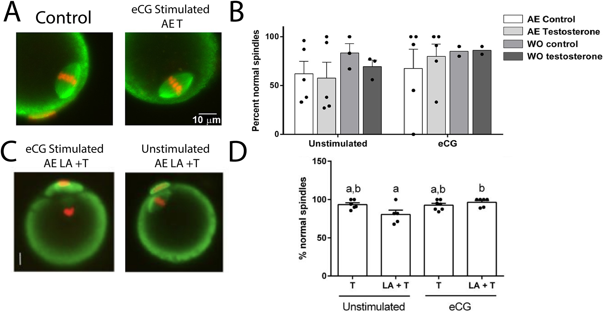
Figure 2. Comparison of Vehicle and T-treated Meiotic Spindle Morphology.

(A) Immunofluorescent stained oocytes from Bartels et al., 2020 showing normal meiotic spindle morphology in oocytes from stimulated T-treated and vehicle mice. (B) T-treated mice produce a comparable number of meiotically competent oocytes to untreated mice regardless of a washout period. (C) Immunofluorescent stained oocytes showing normal meiotic spindle formation regardless of stimulation from Godiwala et al., 2023. (D) Mice treated with T or leuprolide + T produced eggs that had morphologically normal spindles after in vitro maturation (Adapted from Bartels et al., 2020; Godiwala et al., 2023).
