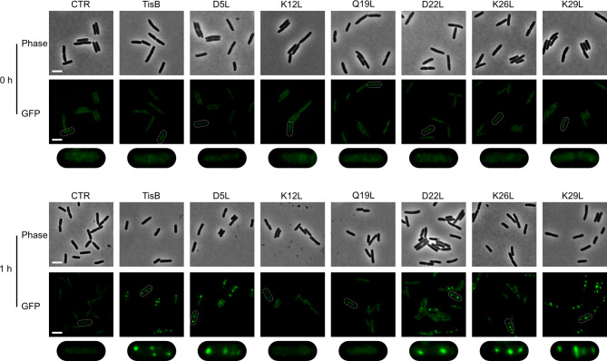
Fig. 2.
TisB-dependent protein aggregation. Reporter strain MG1655 ibpA-msfGFP, harboring p0SD-tisB (TisB) and variants with different amino acid substitutions, were treated with L-ara (0.2%) during exponential phase for one hour. An empty pBAD plasmid (CTR) was used as control. Pre- and post-treatment samples were analyzed by microscopy. Phase contrast (phase) images are displayed together with corresponding fluorescence images (GFP). White bars represent a length scale of 2 μm. The area surrounding a single cell observed in the GFP image (white dashed line) is magnified and shown below the original images for closer inspection.

