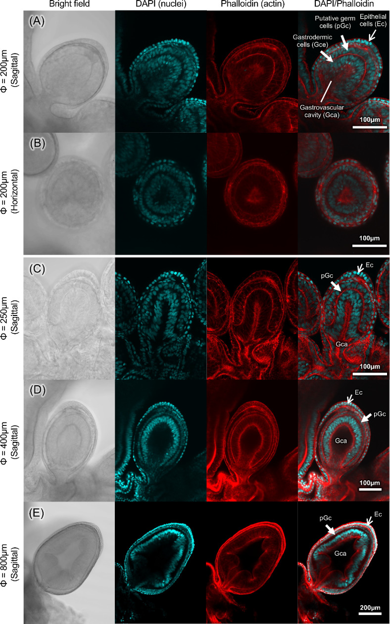
Fig. 2.

Histology of gonophore in various developmental stages of Physalia utriculus. Sagittal section of a gonophore with a major axis diameter of 200 μm (A). Horizontal section of a gonophore with a major axis diameter of 200 μm (B). Sagittal section of a gonophore with a major axis diameter of 250 μm (C), 400 μm (D) and 800 μm (E). From left to right: brightfield, DAPI fluorescence image, phalloidin fluorescence image, and DAPI/phalloidin stacked image. DAPI signals targeting host nuclear DNA (blue), and fluorescent phalloidin signals targeting muscular actin filaments (red), respectively. Epithelial cells (Ec), gastrovascular cavity (Gca), gastrodermic cells (Gce) and putative germ cells (pGc) were seen in every developmental stages.
