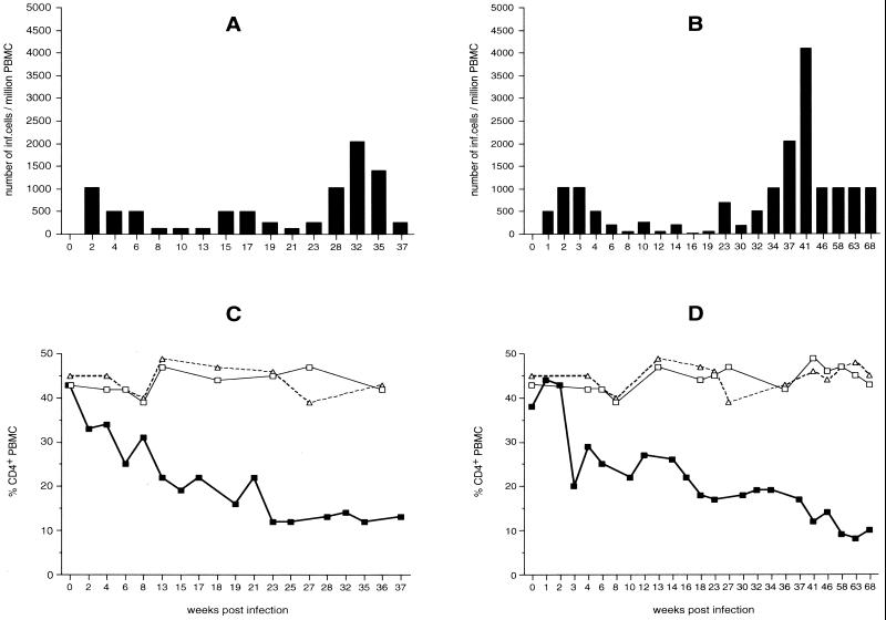
FIG. 3.
Virus load (A and B) and changes in CD4+ lymphocyte populations (C and D) of rhesus monkeys WT (A and C) and L52 (B and D) after infection with molecularly cloned SIVF359. A progressive decline of CD4+ T lymphocytes in infected rhesus monkeys (WT and L52) (▪) compared to control monkeys 8637 (□) and 8711 (▵) is seen.

