FIGURE 8.

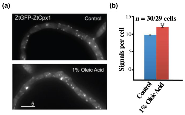
ZtCpx1 is localized to peroxisomes. (a) Zymoseptoria tritici cells expressing ZtGFP‐ZtCpx1 after integration of pCZtGFPCpx1 in to the succinate dehydrogenase locus. 2D‐deconvolved maximum projection of a z‐axis stack, adjusted in brightness, contrast and gamma settings. Bar: 5 μm. (b) Quantitative analysis of peroxisome number per cell. Cells were treated with 1% oleic acid for 1.5 h. Data are from three independent experiments (n = 30/29 cells) and are presented as mean ± SEM. ** indicates significant difference at α = 0.001, Student's t test. Treatment with 1% oleic acid increased the number of fluorescent signals. This strongly indicates that ZtGFP‐ZtCpx1 is localized in the peroxisomes.
