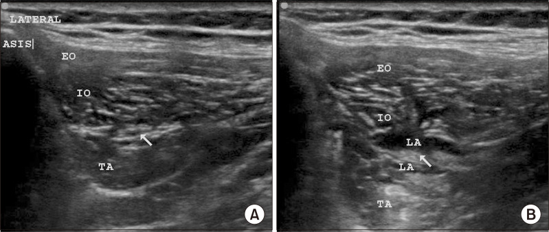
Fig. 2.
Scanning technique for iliohypogastric-ilioinguinal nerve block. (A) The figure shows the sono-anatomy of the block with ilioinguinal/iliohypogastric nerves (the white arrow indicates the fascial plane between IO and TA where the nerves typically travel). (B) The figure shows the local anesthetic (LA) spread around the nerves (marked with a white arrow). ASIS: anterior superior iliac spine, EO: external oblique, IO: internal oblique, TA: transverse abdominis.

