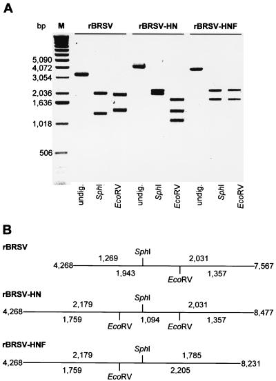
FIG. 2.
Demonstration of marker restriction sites in the genomes of rBRSV, rBRSV-HN, and rBRSV-HNF, confirming the identity of the recombinant viruses. (A) RT-PCR with primers encompassing the glycoprotein genes was performed on RNA of infected cells, submitted to restriction analysis, separated on a 0.8% agarose gel, and stained with ethidium bromide. M, 1-kb DNA ladder (Gibco BRL); fragment size is indicated in base pairs (bp). (B) Schematic diagram of amplified PCR products, with horizontal lines representing the amplified RT-PCR products and vertical bars representing the respective restriction sites. The sizes (in nucleotides) of the resulting DNA fragments are indicated above and below the lines. The positions of the fragments in the genomes of the parental or chimeric virus are indicated on the left and on the right.

