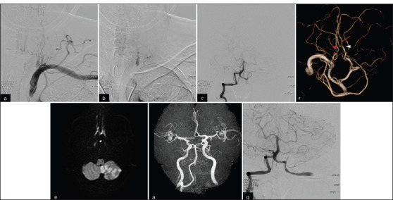
Figure 2:

(a, b) Left subclavian artery angiography shows severe stenosis of the left VA with stagnated distal blood flow and collateral flow from the deep cervical artery to the V2 segment. (c) Right VA angiography reveals retrograde flow to the left V4 segment, which extended to the left posterior inferior cerebellar artery (PICA), and a to-and-flow appearance in the intracranial VA. (d) Three-dimensional rotation angiography reveals a bihemispheric PICA. White arrowhead, the supratonsillar segment of the left PICA; red arrow, the left hemispheric branch; red arrowhead, the right hemispheric branch. (e,f) Follow-up magnetic resonance (MR) imaging reveals enlarged cerebellar infarction, and MR angiography shows poorly visualized left VA. (g) Repeated DSA shows poor filling of the left PICA and proximal translocation of the to-and-flow appearance.
