
Figure 3:
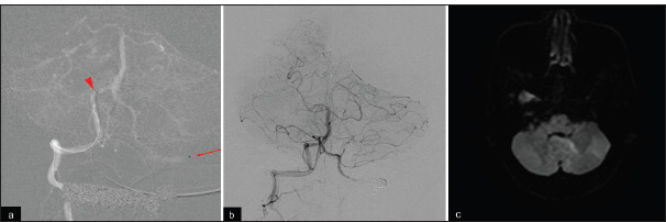
(a) An intermediate catheter (arrowhead) and microcatheter (arrow) were introduced into the right V4 and left V3 segments, respectively. (b) Parent artery occlusion with coils was performed in the left extracranial vertebral artery. (c) Postoperative magnetic resonance imaging reveals no new ischemic infarctions.